Key benefits of Varjo
- Explore complex biological structures and their properties in the highest-fidelity VR
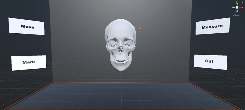
- Bring medical imaging data to VR, AR and XR and ‘dive’ into patients’ bodies
- Accomplish more effective procedures and treatments
- Move freely in a 3D virtual model and focus on the demanding points
- Concentrate on patient data without having to use a mouse or other hand-held controllers
From 2D to the ability to “dive into patients’ bodies”

Digital imaging is an essential part of modern medicine. Magnetic resonance imaging, computed tomography, cone beam computed tomography, and ultrasound imaging provide information for diagnosis and treatment planning.
However, the information technology for producing and processing these images is currently based on two-dimensional slice images traditionally used in the field, although the tissues and structures examined are three-dimensional. And even for specialists, the interpretation of the images is complicated, error-prone, and requires a lot of training and special skills.
Immersive technologies bring completely new possibilities to the field. Varjo is proud to be part of Tampere University’s two-year research project to revolutionize medical imaging. The aim of the project is to allow the representation of complex biological structures and their properties in high-fidelity virtual reality.
Varjo’s human-eye resolution VR devices are a turnkey solution, as they enable more realistic 3D modelling and realistic visualization of medical images inside virtual reality. Ultimately, this helps accomplish more effective procedures and treatments.

High demand for better technical solutions
“Human imaging must be able to represent complex biological structures and their properties. New VR technologies like Varjo make this more effective, as you can see and experience 3D content in human-eye resolution,” says Professor, Head of Computer-Human Interaction Roope Raisamo from Tampere University.
“For example, bringing imaging data to virtual reality enables doctors to ‘dive’ into patients’ bodies. They can move freely in a three-dimensional virtual model and focus on the demanding points. At the same time, haptic feedback improves the understanding of tissues and their properties. As a result, doctors can concentrate on patient data without having to use a mouse or other hand-held controllers. With virtual reality, treatments can be speeded up, made more effective, and their quality improved.”
Problems in medical workflows are a key reason for rising healthcare costs globally, and there’s a high demand for better technical solutions. This two-year research project will contribute to solving many of these problems. In the case of a single patient, immersive technologies can be helpful firstly in diagnostics, then in planning the surgical procedure, and finally during the operation. In the bigger picture, virtual and mixed reality technologies can offer improvements and insight for quality control, educational methods, and scientific research.
Learn more
- Read the full press release by Tampere University
- Learn more about using Varjo’s VR/XR for medical purposes
- Contact your Varjo experts


